
Neck & Upper Limb Tests

Cartoid & Vertebral Arteries
Upper Limb Arteries
Fistula Mapping & Monitoring
Transcranial Arteries
Upper Limb Veins
Giant Cell Arteritis (GCA) Study
Thoracic Outlet Study
Carotid & vertebral arteries
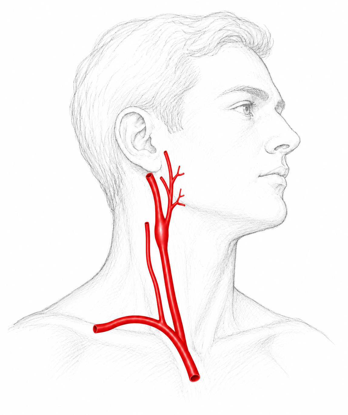
Procedure
A duplex of the extracranial carotid, vertebral and subclavian arteries to identify and quantify plaque, vertebral “steal” (reversal of flow direction), vessel occlusion, and wall irregularities (i.e. fibromuscular dysplasia). Less common findings include aneurysms, vasculitis (Takayasu’s and Giant cell arteritis), and carotid body tumours.
Indications
Stroke or TIA, bruit, visual disturbance, lipid dysregulation, pre-op assessment, known disease monitoring.
Transcranial arteries
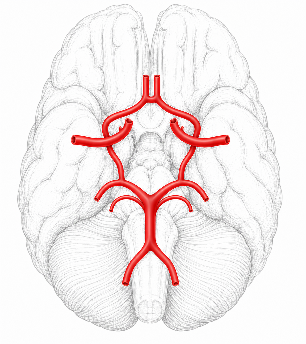
Procedure
A duplex of the cerebral arterial circle to assess for patency, flow direction and stenosis.
Indication
This is usually performed when the patient has significant extra-cranial carotid disease (i.e >80% stenosis).
Giant Cell arteritis (GCA) study
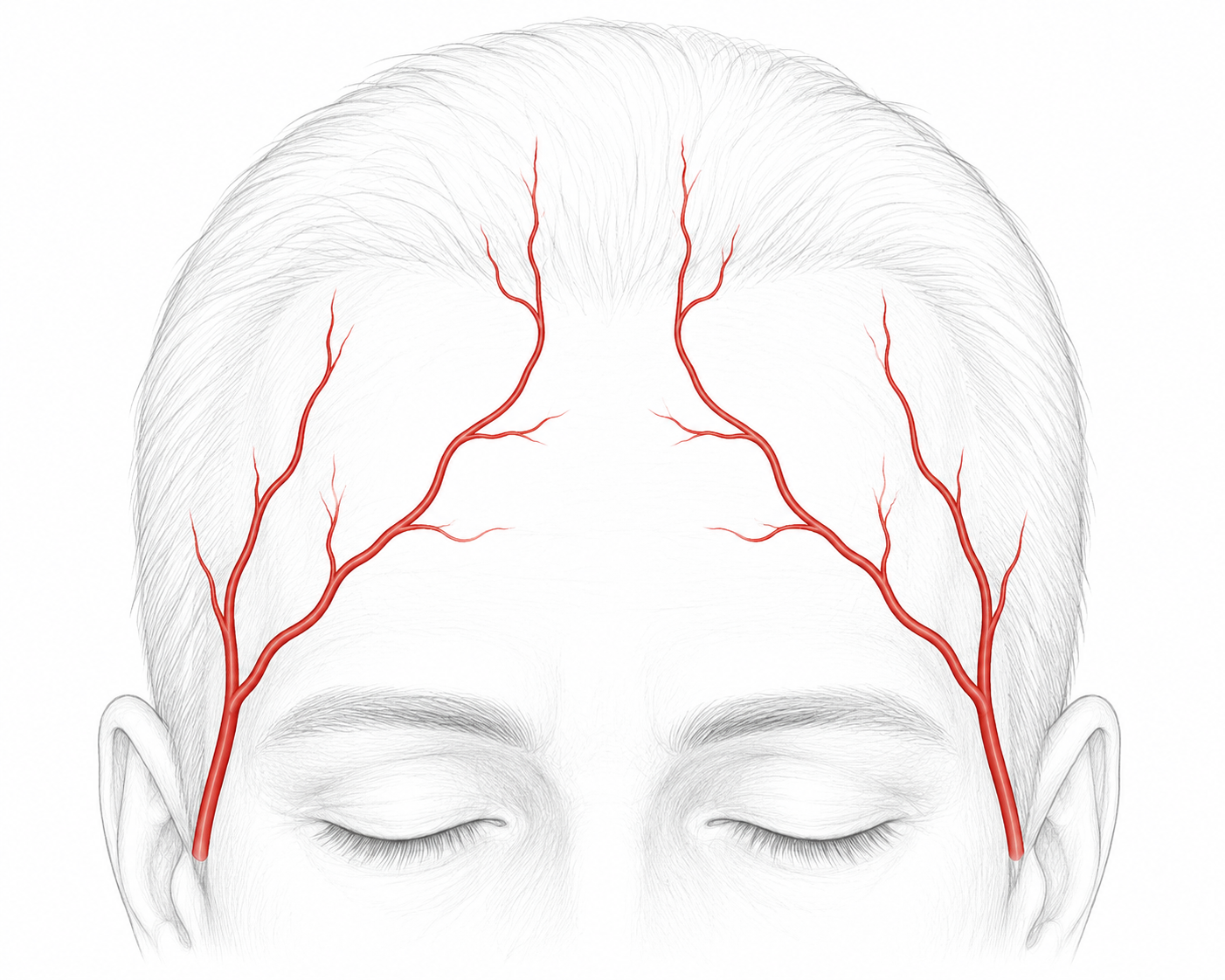
Procedure
A duplex to assess for the presence of arteritis (seen as intimal thickening) in the extracranial carotid, vertebral, subclavian, axillary, superficial temporal and occipital arteries. Ultrasound is very effective in identifying segmental GCA “skip-lesions”, which can be missed with biopsy.
Indications
New or atypical temporal headache, scalp tenderness, visual disturbance, jaw claudication, polymyalgia rheumatica.
Upper limb arteries
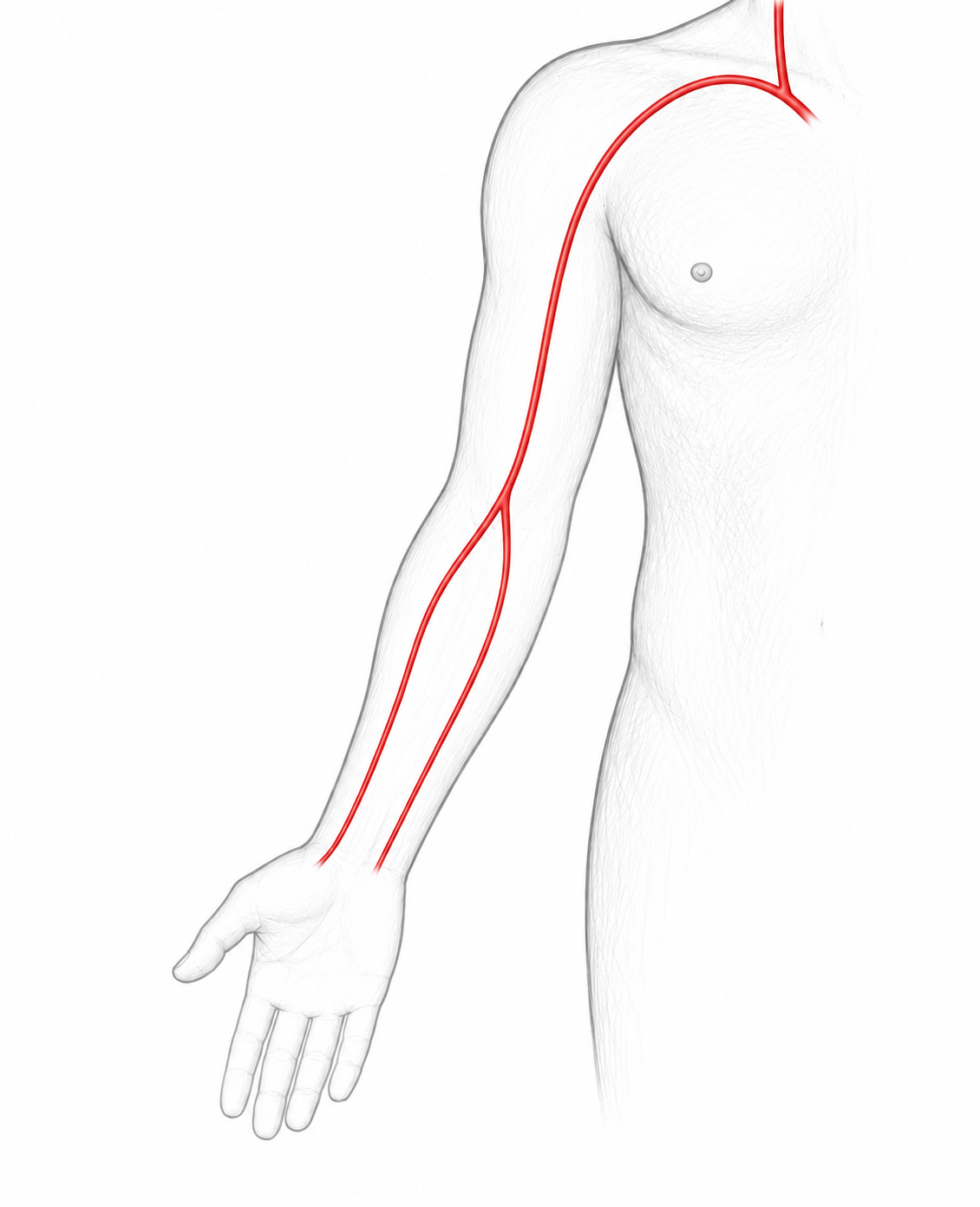
Procedure
A duplex of the subclavian, axillary, brachial, radial and ulnar arteries to assess for plaque, thrombus, aneurysms and pseudoaneurysms.
Indications
Ischemic digits, pulsatile lump, asymmetric brachial pressures, pre-operative mapping.
Upper limb veins
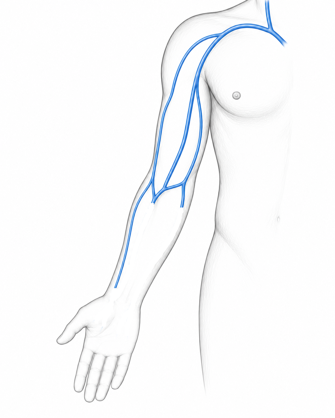
Procedure
A duplex of the subclavian, axillary, brachial, cephalic and basilic veins to assess for thrombosis and measure its extent.
Indications
Pain or palpable lump in arm, acute arm swelling, pulmonary embolism, known thoracic outlet compression.
thoracic outlet study
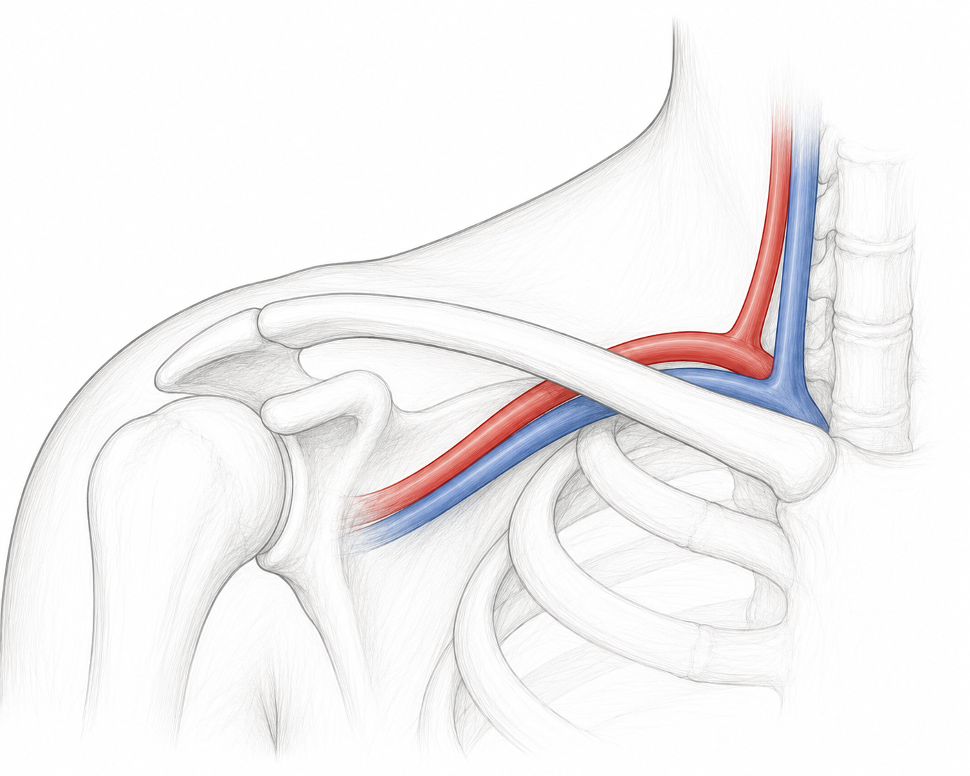
Procedure
A functional duplex to assess the subclavian and axillary arteries and veins for dynamic compression. The patient will be required to perform various provocative manoeuvres during this scan.
Indications
Position-dependent sensory changes such as pain, tingling or numbness in the hands, cyanosis (colour change), prominent chest-wall veins.
fistula mapping & monitoring
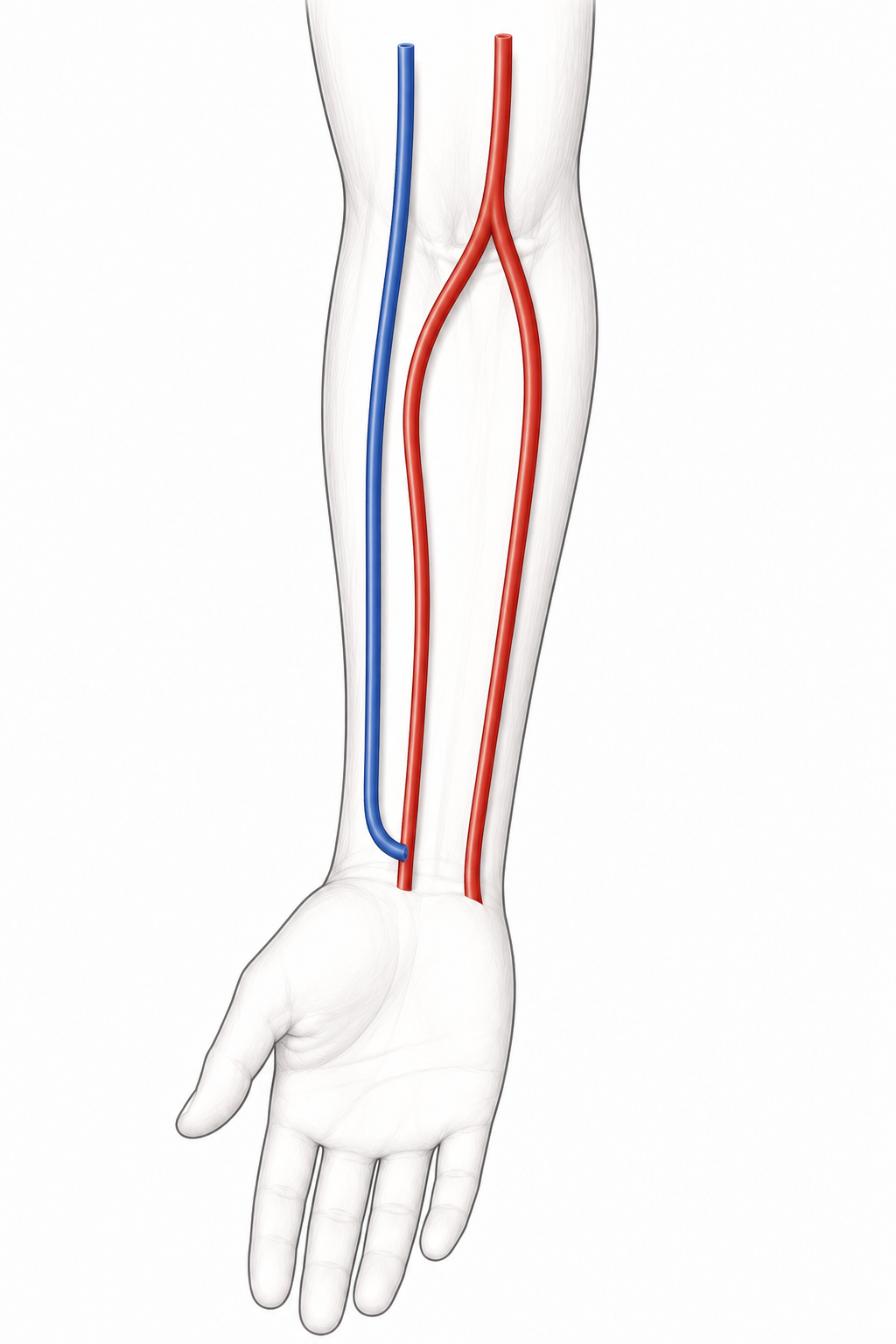
Fistula mapping uses ultrasound to assess a site (usually the non-dominant arm) for suitability to create a fistula for dialysis access. A detailed report is formulated including diameters of all relevant arteries and veins, so the specialist can make an informed decision regarding the appropriate site for the fistula.
Once a fistula is formed it is common for patients to have regular surveillance with ultrasound to ensure it is functioning well, and to monitor for pathological changes that require intervention.